T and B lymphocyte life cycle - naive lymphocytes
The vast majority of lymphocytes do not receive the appropriate survival and maturation signals during their development in the primary lymphoid organs and die by apoptosis. Only around 2% reach full maturity and are released into the circulating blood and lymphatic systems as naive lymphocytes, proudly displaying newly constructed antigen specific receptors. What do they do next and where will they end up in the body?
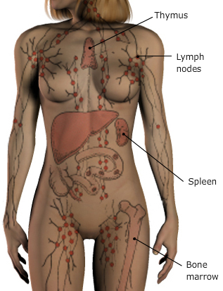
Naive lymphocytes are now ready to respond to antigens and as secondary lymphoid organs serve as antigen traps, naive lymphocytes migrate to these organs in search for antigens. As you can see in the graphic, secondary lymphoid tissues are distributed all over the body like a suit of armour, only on the inside! But how do naive lymphocytes know to go there? Lymphocytes move around the body by squeezing out of blood vessels and into different tissues. Blood vessels are influenced by the tissues they are permeating and this results in sections of blood vessels being coated on the inside with different proteins. Lymphocytes also display different surface molecules at different stages of their life cycle, for example, naive T cells express a lot of the chemokine receptor called CCR7, but this receptor is lost after the T cell has become antigen experienced. See if you can figure out why the naive T cell in this video homed to the lymph node?
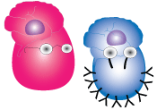
It wasn't just the MIP-3β interacting with CCR7 that pulled the naive T cell into the lymph node but also other adhesion molecules on the high endothelial venule (HEV). Naive B and T cells circulate as resting cells, but when a naive lymphocyte finds its antigen it becomes activated and as we saw in the video, it starts to divide. A population of cells descending from a single activated B or T cell is called a clone. An immune response is called monoclonal if the antigen stimulates a single B or T cell to divide. Usually an antigen stimulates more than one B or T cell and this produces a polyclonal response. You can see how a polyclonal response could come about in the graphic where two different B cells are recognising different parts of the same antigen. When activated, they will both start dividing and producing antibodies, resulting in the polyclonal response. A mixture of antibodies produced by several B cells means that many different antibodies are coating the antigen which makes it more readily detected and cleared by garbage-disposal phagocytic cells.